Friday, October 27, 2017

e-portfolio sixth week

      During the lab this week, I had done 3 experiments which is experiment 6( preparation of bacterial smear), experiment 7( simple staining) and experiment 8 (negative staining).All the experiment we did used gram-negative bacteria. For the bacterial smear we have done bacterial smear  to be used for the simple staining and negative staining experiment. The bacteria we used are Staphylococcus aureus, Bacillus cereus, Escherichia coli, Micrococcus Luteus and Bacillus subtilis. There are two type of bacterial smears which are the smears from a broth medium and the smears from a solid medium. The bacterial smears we done are the smears from solid medium which is the agar plate. The process of carrying out bacterial smear follow the following procedures. First, clean the glass slides by flaming it. Then, using a loop, place one to two loops of water on each slides. Then, with a sterile loop, we touch the entire loop to the culture and emulsify the cells in water on all the slides respectively. Next, we allow the slides to air-dry completely by placing them on the bench.Finally, we heat fix the preparation.

   We did simple staining for Escherichia coli, Bacillus cereus and Staphylococcus aureus.The stains we used are the carbolfuchsin, methylene blue and crystal violet. We did it in a group and each bench must prepare two sets of the simple staining of the bacteria for each different stains. We prepared 18 slides in total. For the procedures of the simple stain, we used the bacterial smears we prepared during experiment 6. First, place a slide on the staining tray and flood the smear with one of the indicated stains, using the appropriate exposure time for each: carbolfuchsin,15-30 second; crystal violet,20-60 seconds; methylene blue, 1-2 minutes. Then, we gently wash the smear with tap water to remove excess stain. During this step, hold the slide parallel to the stream of water, in this way, I can reduce the loss of organisms from the preparation. Next, we blot dried the slide by using bibulous paper. We did the procedures for all the organisms by using all three different type of stains for each organisms.

 For negative stains, the background is stained dark while the bacteria remains colourless. This is because the stain we used are nigrosin which acidic stain which negatively charged chromogen repel with the gram-negative bacteria. So, the bacteria resists staining and remained colourless. The bacteria we used are Micrococcus luteus, Bacillus cereus and Staphylococcus aureus. The procedures of negative staining is as followed. First, we placed a small  drop of nigrosin close to one end of a clean slide. Then, by using aseptic technique, we placed a loopful of inoculum from the M. luteus culture in the drop of nigrosin and mix. We then placed a slide against the drop of suspended organisms at 45 degree angle and allow the drop to spread along the edge of the applied slide. Next, we push the slide away from the frop of suspended organisms to form a thin smear. We air-dried the slides. Heat fix should not be done for negative staining. We repeat the steps 1-4 for slide preparations of the remaining cultures, We then examined all the slides under high power and oil immersion and recorded the result in the Lab report.


Simple staining using crystal violet, methylene blue and carbolfuchsin and negative staining using nigrosin for E. coli


The slide of E. coli of carbolfuchsin (simples staining)
The slide of E. coli for negative staining
The slide of S.aureus for simple staining (crystal violet)
 
 The slide of B.cereus using methylene blue (simple staining)


Saturday, October 14, 2017

eportfolio-week 5

During this week,we have done an experiment which is experiment A, which is the microscopic measurement of microorganisms. In this experiment, I learnt to calibrate on ocular micrometer. The ocular micrometer is inserted into the ocular lens.Then, I observe the ocular lens with the calibration.Then, I put the stage micrometer with 0.01mm on the stage.Then, I superimposed teh ocular micrometer over the stage micrometer.I calculate the calibration factor for the magnification of 100x, 400x, and 1000x and one ocular division for each  magnification.One ocular division on ocular micrometer in mm is calculated by using the formula : (known distance between two lines on stage micrometer/number of divisions on ocular micrometer). Then, I observed the prepare sides of yeast cells, protozoa, and bacterial cocci and bacilli under the all the magnification and calculate the number of ocular division for each microorganisms. Then, I calculated the length of  organism by using the formula :(number of ocular divisions occupied x calibration factor for one ocular division)

Micrograph of yeast cell with calibration

Saturday, October 7, 2017

e-portfolio fourth week (experiment 4 and 5)

Experiment 4:Microscopic examination of stained cell preparation
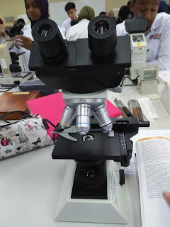
During the fourth experiment, I have the chance to use the compund light microscope.We are told how to handle the microscope carefully.When holding the microscope,we need to put one hand at the arm of the microscope while the other hand at the base of the microscope.Then,we put the mciroscope at our bench.We were given some prepared slides of Staphylococcus aureus, Bacillus subtilis, Bacillus cereus, slide  of blood smear and slides of Saccharomyces cerevisiae.We observe the slide starting by using magnification of 40x,followed by 100x, 400x and 1000x(oil immersion).

Pictures of compound light microscope

Slide of Staphylococcus aureus (high power)
 Slide of Saccharomyces cerevisiae (high power)
 Slide of Bacillus subtilis (high power)
 Slide of Bacillus cereus (high power)
 Slide of Saccharomyces cerevisiae

 Slide of red blood cell


Experiment 5:Microscopic Examination of Living Microorganisms Using a Hanging-Drop Preparation or a Wet Mount
In this experiment,I learnt how to prepare a hanging-drop and wet mount.To prepare the hanging-drop,I have to spread a ring of petroleum jelly around the concavity of the depression slide.Then,I place of loopful of the bacterial culture in the center of the coverslip. Then,I lower the depression slide,with the concavity facing down,onto the coverslip and press gently to form a seal.Finally,I turn the hanging-drop preparation over so that the culture drop adheres to the coverslip.For the wet mount,
I use the cotton swab to apply a thin layer of petroleum jelly along the edge of the four sides of a coverslip.Then,by using the aseptic techniques,I place a loopful of the culture in the center of the coverslip.After that,I place a clean glass slide over the coverslip and press the slide gently to form a seal between the slide and coverslip.Next,I turn the wet mount preparation slide over so that the culture drop adheres to the coverslip.We did the hanging drop and wet mount preparation for the broth cultures of P.aeruginosa, B.cereus,S.aureus,,P.vulgaris,hay infusion and pond water.I observe it under high power and oil immersion and record the observation in a table.
Slide of Bacillus cereus
 Slide of S.aureus


Eportfolio week 13

During the lectures, we have a quiz section where each groups prepared some questions. There are some questions asked such as what will happ...