Sunday, November 26, 2017

eportfolio week 10

Experiment 14 and 15
This week I carry out experiment 14 (Physical Factors: Temperature) and experiment 15 ( Physical Factors: pH of The Extracellular Environment. In experiment 14, the procedures of doing the experiment were as followed. We first scored the underside of all plates into four quadrants with a glassware marker. We labelled each section with the name of the test organism to be inoculated. We included the temperature of incubation (4°C, 20°C 37°C or 60°C) when labelling the cover of each plates. Then, we aseptically inoculated w=each of the plates with E. coli, B. stearothermophilus, P. aeruginosa and S. marcescens by means of a single line of inoculation of each organism in its appropriately labelled section. Then, we appropriately labelled the four Sabouraud broth tubes, including the temperatures of incubation as indicated above. We then gently shook the S. cerevisiae culture to suspend the organisms. We aseptically added one drop of the culture into each of the four tubes of broth media by using a sterile Pasteur pipette. All the plates and the broth cultures were then incubated in an inverted position abd at each of the four experimental temperatures. (4°C, 20°C 37°C or 60°C) for 24 to 48 hours. We then observed all the growth of the culture and classified the organisms as psychrophiles, mesophiles, facultative thermophiles or obligate thermophiles respectively at the next day. This experiment enabled me to determine whether the optimum growth temperature is also the ideal temperature for enzyme-regulated cell activities such as pigment production and carbohydrate fermentation. I also learnt about the diverse growth temperature requirements of bacteria. 

In experiment 15, the procedures of the experiment were as followed. First, I inoculated a series of the appropriately labelled TSB (12 Trypticase soy broth) tubes of media, pH values of 3, 6, 7, and 9 with E. coli by adding 0.1 ml of the saline culture to each.  I repeated the step just now for the inoculation of A. faecalis and S. cerevisiae by using a new sterile pipette each time. Finally, we incubated the A. faecalis and E. coli cultures for 24 to 48 hours at 37°C and the S. cerevisiae cultures for 48 to 72 hours at 25°C. We used the spectrophotometer to determine the absorbance of all cultures and record the readings in the Lab Report.

Sunday, November 19, 2017

Eportfolio week 9

This week I have done two experiment which are experiment 12 and experiment 13. Experiment 12 is on Nutritional Requirements: Media for the Routine Cultivation of Bacteria while Experiment 13 is on the Use of Differential, Selective, and Enriched Media. For experiment 12, the procedures are as followed. We first inoculate the microorganisms (E. coli, A. faecalis and S.mitis) to different media ( yeast extract broth, nutrient broth, glucose broth and inorganic synthetic broth in four test tubes respectively by using Mohr pipette. After inoculate the broth culture and incubate the culture for 24 to 48 hours, we use the spectrophotometer to measure the optical density readings with the wavelength of 600nm. First, we measure the blank of culture, then we only measure the optical density for different media containing different microorganisms.

For experiment 13, we do the experiment by following the procedures. We label the cover of the plate properly and divide each of the Petri dishes into the required number of section by marking the bottom of the dishes. Each section are labelled with the name of the organism to be inoculated. Using aseptic technique, we inoculate all plates, except the blood agar plate, with the designated organisms by making a single line of inoculation of each organism in its appropriate section. Using aseptic techniques, we inoculate the blood agar by making a single line of inoculation. On completion of each single line of inoculation, we use the inoculating loop and make three or four stabs at a 45 degree angle across the streak.

We inoculate each of the different media with the following:
a) Phenylethyl alcohol agar: E.coli, S. aureus, and E. faecalis.
b) Crystal violet agar: E. coli, S.aureus, and E. faecalis.
c) 7.5% sodium chloride agar: S. aureus, S. epidermidis and E. coli
d) Mannitol salt agar: S. aureus, S.epidermidis, E. aerogenes. and E.coli
e) MacConkey agar: E. coli, E. aerogenes, S. typhrimurium, and S. aureus
f) Eosin-methylene blue agar: E. coli, E. aerogenes, S. typhimurium, and S. aureus
g) Blood agar: E. faecalis, S. mitits, and Streptococcus pyogenes.

The phenyethyl alcohol agar plate was incubated in an inverted position for 48 to 72 hours at 37 degree celcius while the remaining plates were incubated in an inverted position for 24 to 48 hours at 37 degree celcius.

The other day, we examine each plate and recorder the result according to the amount of growth along line of inoculation, appearance of the growth and the change in appearance of the medium surrounding the growth.

The purpose of this experiment is to understand which medium is suitable to culture which microorganisms.
The picture of the spectrophotometer

The picture of the culvate and the nutrient broth

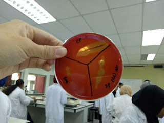
 The picture of the blood agar with microorganisms after incubation

 The picture of phenyethyl alcohol agar plate with microorganisms after incubation

E-portfolio week 8

This week I have my hands on test on streak plate and simple staining. Then, I did an experiment on capsule stain and spore stain. For spore stain, the procedures for the experiment were as followed. Individual smears were made in usual manner by using aseptic technique on two clean glass slides. The smears were allowed to air-dry and heat fix in the usual manner. Then. the smears were flooded with malachite green and placed on top of a beaker of water sitting on a warm hot-plate, allowing the preparation to steam 2 to 3 minutes. The slides were then removed from the hot plate, cooled and washed under running tap water. Next, the slides were counterstained with safranin for 30 seconds and washed with tap water. Finally, the smears were blotted dry with bibulous paper and examined under oil immersion. The organisms used are Bacillus cereus and Clostridium sporogenes

For capsule stain, I did the following procedures.The procedures of the experiment were as followed. One glass slides was obtained and several drops of crystal violet stain were placed on the slide. By using aseptic technique, three loopfuls of Klebsiella pneumonia were added to the stain and mixed gently with the inoculating loop. Then with a clean slide, the mixture was spreaded over the entire surface of the slide to create a very thin smear and the slide was let stand for 5 to 7 minutes. The smear was allowed to air-dry. The smears were washed with 20% copper sulphate solution. Finally, the smears were blotted dry and examine under oil immersion. Step 1 to 5 was repeated for Enterobacter aerogenos.
            The procedures of negative staining were also done to observe the capsule of the organisms by using nigrosin. The procedures for negative stain were as followed. First, a small drop of nigrosin was placed close to one end of a clean slide. Then, a loopful of inoculum from the Klebsiella pneumonia culture was placed in the drop of nigrosin by using aseptic technique and mixed.  Next, a slide was placed against the drop of suspended organisms at 45° angle and the drop was allowed to spread along the edge of the applied slide. The slide was then pushed away from the drop of suspended organisms to form a thin smear. Steps 1 to 4 were repeated for slide preparation of the remaining cultures. The slides were then examined under oil immersion.

Slides of the microorganisms for spore stain,capsule stain and negative stain


Micrograph of Bacillus cereus under 1000X magnification

Micrograph for capsule staining under 1000X for Enterobacter aerogenes.


Saturday, November 4, 2017

eportfolio basic technique week 7

Experiment 9 and 10
This week, the experiment that I have done is the gram-staining and acid-fast staining. Gram staining is a differential staining which determine whether the bacteria is gram positive or gram negative. All the bacteria is negatively charged. Gram staining differentiates the bacteria based on the cell wall which is the thickness of the peptidpoglycan. Gram positive bacteria has a thicker cell wall. Therefore, the crystal violet can be retained in the cell wall. Gram negative bacteria has a thinner cell wall. Therefore, it only retains the counterstain safranin. The procedure of gram staining is as followed. First, we gently flooded the smears with crystal violet and let stand for 1 minute. Then, we washed it with tap water. Next, we flooded the smears with Gram's iodine mordant and let stand for 1 minute. The smear is then later decolorized with 95% ethyl alcohol. The reagent was added drop by drop until the alcohol runs almost clear,showing only a blue tinge. Then, we gently washed the smear with tap water and counterstained with safranin for 45 seconds. It was gently washed again with tap water. The smear was then blotted dry with bibulous paper and examined under oil immersion. The gram staining were done for cultures of E. coli, S. aureus , B. cereus and the mixture of S.aureus and E. coli.

The next experiment which is experiment 10 is acid-fast staining. Acid-fast staining shows postive result for bacteria which has a thick, waxy wall that makes penetration by stains extremely difficult. The postive result is the smear of the bacteria will become pink in colour while negative results is the bacteria smear become blue in colour. The procedure of acid-fast stain is as followed. Smear of Mycobacterium smegmatis , Staphylococcus aureus and the mixture of both bacteria are prepared. The smears are then flooded with carbol fuchsin and placed over a beaker of water on a warm hot plate, allowing the preparation to steam for 5 minutes. Next, the smears were washed with tap water and heat slides must be cooled prior to washing. Then, decolorize the smears with acid-alcohol, adding the reagent drop by drop until the alcohol runs almost clear with a slight red tinge. The tap water was used to wash the smear. Finally, blot dry the smear with bibulous paper and the slides were examined under oil immersion.
The slides of gram-staining and acid-fast stain

 The slides of Bacillus cereus under oil immersion
 The slides of the mixture of S. aureus and E. coli
The slides of M. smegmastis under oil immersion


Eportfolio week 13

During the lectures, we have a quiz section where each groups prepared some questions. There are some questions asked such as what will happ...